


Edentulous scanning is one of the most challenging applications in digital dentistry. In tooth-supported cases, scanners can rely on cusps, grooves, line angles, and other clear landmarks to maintain tracking and rebuild a stable 3D model. In edentulous arches, those natural reference points are largely missing.
That changes everything.
Clinicians often find that a scanner that performs very smoothly in single crowns or short-span restorative work feels far less forgiving when scanning a full edentulous arch. Tracking may drift, soft tissue may distort, and the digital model may look complete at first but prove less reliable when used for denture design, implant planning, or guided workflows.
The good news is that edentulous scans can be made much more reliable with the right approach. Success depends less on “scanning faster” and more on understanding what makes edentulous anatomy difficult for scanners in the first place.

Why Edentulous Cases Are More Difficult to Scan
The main issue is the lack of stable geometry.
In dentate scans, the scanner continuously recognizes tooth anatomy and uses it as a reference for stitching frames together. In edentulous scans, the software has to track across broader, smoother, and more repetitive soft tissue surfaces. That makes it easier for the scan to lose positional confidence over distance.
Edentulous arches also introduce other challenges:
-
compressible soft tissue that changes under pressure
-
limited visual landmarks in flat ridge areas
-
saliva pooling in vestibular regions
-
mobile lips, cheeks, and tongue affecting the scan field
-
greater risk of cumulative stitching error over long spans
This is why edentulous scans often fail not because the scanner “cannot scan soft tissue,” but because the workflow becomes less stable once tracking and tissue control start to break down.
Start with the Right Expectations
One reason edentulous scanning becomes frustrating is that many clinicians approach it like a normal crown scan—just with more surface area. In reality, it is a different scanning task with different priorities.
The goal is not only to capture anatomy. It is to maintain a stable digital path over tissue that is inherently less structured and more variable than teeth.
That means the workflow should prioritize:
-
tracking stability
-
tissue visibility
-
consistent scanning path
-
minimal distortion from movement or compression
Once those priorities are clear, technique becomes much easier to refine.
Control the Soft Tissue Environment First
In edentulous cases, soft tissue management matters more than speed.
Before scanning:
-
dry the arch gently but thoroughly
-
reduce pooled saliva, especially in the vestibular area
-
retract lips and cheeks enough to expose the functional anatomy without stretching tissue unnaturally
-
avoid pressing the scanner tip into soft tissue, which can distort the surface
Even slight tissue compression can change the shape of the recorded anatomy. In a denture or full-arch workflow, that can affect fit, border design, and overall reliability.
A clean and stable field is often more important than trying to complete the scan quickly.
Use a Deliberate, Structured Scan Path
Edentulous arches are much less forgiving when the scan path is inconsistent. Random movement, repeated jumps between areas, or broad sweeping rescans make it easier for the software to lose spatial orientation.
A more reliable approach is to use a simple and repeatable sequence:
-
begin in an area with the strongest visible morphology
-
move in a continuous path without abrupt changes in direction
-
cover the crest first, then transition gradually to buccal and lingual surfaces
-
avoid leaving large unscanned gaps that must be reconnected later
The exact path may vary by operator or scanner, but consistency matters more than personal preference. A controlled path reduces stitching drift and gives the software a better chance to maintain model stability over the full arch.
Pay Attention to Scan Range and Tracking Tolerance
Edentulous scans often involve awkward angles, posterior reach limitations, and surfaces that do not provide strong visual reference. That makes scanner tolerance more important.
In these cases, a scanner with a more forgiving effective scan range can help the operator maintain tracking without constantly searching for the perfect distance or angle. This becomes especially useful when scanning the distal ridge, deeper vestibular areas, or less accessible posterior anatomy.
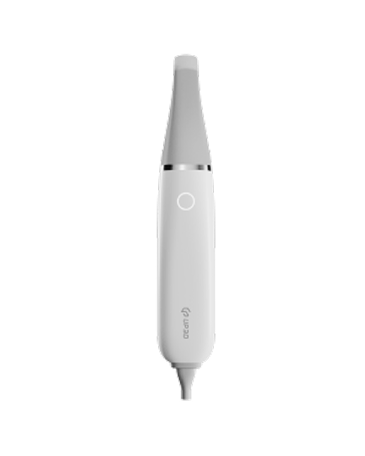
That is one reason clinicians often appreciate deeper scan capability in challenging workflows. Scanners such as the UP610, for example, are often valued not only for speed, but for being more adaptable in situations where anatomy is harder to capture cleanly and consistently.
The point is not that scan depth solves every edentulous challenge. It is that better scan tolerance can reduce interruptions in cases where ideal positioning is rarely possible.
Avoid Overscanning and Random Corrections
When a scan begins to drift, the instinct is often to go back and “fix” the problem by scanning over the same areas repeatedly. In edentulous cases, that usually makes things worse.
Repeated rescanning can:
-
introduce overlapping soft tissue data
-
create conflicting geometry
-
confuse the stitching algorithm
-
increase the risk of distortion over long spans
If the scan clearly loses stability, it is often better to stop and rescan a smaller section carefully than to keep layering more data onto an already unstable model.
Reliable edentulous scanning depends on clean data, not excessive data.
Use Landmarks When Available
Some edentulous cases offer more natural reference than others. If there are remaining landmarks—such as retromolar pads, distinct ridge contours, or implant scanbodies—these areas can help stabilize the scan.
In implant-related edentulous cases, scanbodies often provide much stronger geometry than surrounding soft tissue. Using them strategically within the scan sequence can improve tracking and overall alignment.
The broader principle is simple: whenever the software has better reference points, stitching becomes more reliable.
Check the Model for Drift Before Moving On
An edentulous scan can look acceptable locally while still drifting globally. That is why visual review is essential before the file moves into design.
Look for signs such as:
-
asymmetry that was not present clinically
-
smooth tissue areas that appear doubled or wavy
-
vestibular regions that seem stretched or unstable
-
posterior areas that no longer align naturally with the rest of the arch
These are often early signs of stitching drift. Catching them before export saves much more time than trying to correct the consequences later in denture design or implant planning.
Why Edentulous Scanning Technique Matters So Much in Digital Denture Workflows
Edentulous scanning is no longer a niche skill. As digital denture workflows continue to expand, clinicians are increasingly expected to capture useful edentulous data at the source.
If the scan is unstable, the problems do not stay at the scanning stage. They continue downstream into:
-
denture base design
-
tissue adaptation
-
bite verification
-
try-in predictability
-
final fit
This is why scan reliability matters so much in edentulous cases. The digital workflow becomes only as stable as the anatomy that was captured at the beginning.
The Best Results Come from a Different Mindset
Clinicians who scan edentulous arches well usually do not treat these cases as “harder versions” of normal scans. They treat them as their own workflow.
That means:
-
slowing down when needed
-
respecting tissue behavior
-
following a structured scan path
-
recognizing when the model is drifting
-
using scanner tolerance to support, not replace, good technique
Once that mindset changes, edentulous scans become much more predictable.
Final Thoughts
Edentulous cases are more difficult to scan because the scanner has less natural geometry to rely on and the tissues themselves are more variable. But difficulty does not mean unreliability.
With good field control, a disciplined scan path, careful handling of soft tissue, and realistic expectations, clinicians can capture edentulous arches much more consistently than many assume. In this workflow, reliability comes from process control—not speed alone.
As digital denture and implant workflows continue to grow, the ability to scan edentulous cases well is becoming an increasingly valuable skill. And in those cases, the scanners that offer stronger tracking stability and better scan tolerance often make the biggest practical difference exactly where scanning becomes most difficult.