


Implant scanbody scanning is one of the most important steps in a digital implant workflow—and one of the easiest places for subtle errors to begin. When the scanbody is captured accurately, the lab receives reliable positional data, the CAD software can identify the implant correctly, and the restoration design starts from a stable foundation. When the scanbody is scanned poorly, the entire workflow becomes less predictable.
What makes this challenging is that scanbody scans often look acceptable on screen, even when accuracy is already compromised. The operator may see a complete model, but the data can still be slightly distorted, incomplete, or misaligned in ways that only become obvious later during design, milling, or seating.
This is why implant scanbody scans fail more often than many clinicians realize—and why improving them requires more than simply “getting the body in the scan.”

Why Scanbody Accuracy Matters So Much
In a standard crown case, a small local scanning issue may affect a margin or contact area. In an implant case, scanbody capture does something more fundamental: it defines the implant position itself inside the digital workflow.
That means the scan is not only capturing shape. It is providing information about:
-
implant location
-
implant angulation
-
rotational orientation
-
relationship to adjacent teeth and soft tissue
If any of this information is incomplete or distorted, the downstream restoration may be designed on a faulty reference. The result can be poor seating, contact discrepancies, screw access misalignment, or loss of passive fit.
That is why scanbody scans deserve much more attention than they often receive.
One of the Biggest Problems: Incomplete Geometry Capture
A scanbody only works when the software can read its geometry clearly and completely.
Many scanbody scan failures begin because the operator captures only the visible outer surface while missing the geometry that the software actually needs for reliable alignment. This often happens when:
-
the scanner angle is too limited
-
tissue or saliva obscures part of the scanbody
-
the operator moves too quickly around the body
-
tracking is lost and re-established inconsistently
A scanbody does not need to look “mostly there.” It needs to be captured with enough definition that the software can match it correctly and confidently.
This is one reason why implant scans can fail even when the model appears visually complete.
Soft Tissue Interference Is More Important Than Many Clinicians Expect
Implant scanbody scans are highly sensitive to soft tissue around the implant site.
If tissue overlaps the base of the scanbody, rebounds during scanning, or is not fully controlled, several problems can occur:
-
the body appears shorter or partially hidden
-
the margin between scanbody and tissue becomes unclear
-
the software receives conflicting geometry
-
scanning around the base becomes inconsistent
Unlike natural tooth scans, scanbody scans depend heavily on clear mechanical geometry. Soft tissue that obscures the lower portion of the scanbody can reduce the scanner’s ability to capture that geometry reliably.
Good tissue control is not optional here—it is part of the accuracy protocol.
Moisture and Reflection Can Disrupt Scanbody Recognition
Scanbodies are not scanned under ideal dry-lab conditions. They are scanned in the mouth, where saliva, light reflection, and difficult access often combine in the same small area.
Moisture can create reflective artifacts, especially when it pools around the base of the scanbody or along nearby restorative surfaces. This can interfere with:
-
edge recognition
-
local stitching stability
-
geometric clarity
The more reflective or contaminated the area becomes, the more the scanner may struggle to maintain clean and readable data.
Even modern scanners with better noise handling still benefit from careful surface drying and controlled field visibility when scanning scanbodies.
Angle and Access Problems Increase Error Risk
Posterior implant cases are especially demanding because the operator often cannot hold the scanner at an ideal angle.
This becomes more difficult when:
-
the patient has limited opening
-
the implant is distal
-
the scanbody is tall or positioned close to adjacent structures
-
the scanner tip cannot approach the area comfortably
When the scanner must work at compromised angles, the risk of incomplete capture increases. This is also where scanner tolerance becomes clinically important. A scanner that requires very strict positioning may be more difficult to use reliably in these situations than one with a more forgiving effective scan range.
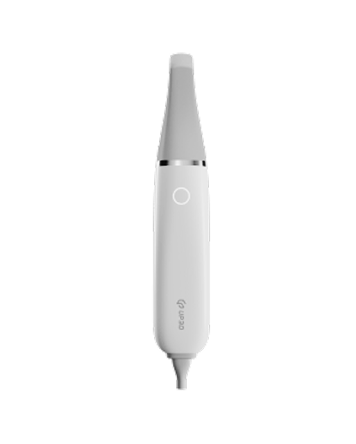
That is one reason scanners with deeper usable scan depth—such as the UP610—are often appreciated in implant workflows, especially where posterior access and angle control are less than ideal. In these cases, practical scanning tolerance can reduce interruption and make scanbody capture more stable.
Scan Path Discipline Matters More Than Speed
Many scanbody errors are not caused by one major mistake. They are caused by unstable scan behavior around a very important structure.
Common scan path problems include:
-
circling the scanbody too quickly
-
scanning it from random directions
-
repeatedly jumping away and back again
-
trying to “fix” the body by layering more and more local data
This kind of overscanning often creates data confusion rather than better accuracy.
A better approach is usually:
-
stabilize the field first
-
approach the scanbody deliberately
-
move around it in a controlled and readable path
-
confirm geometry before continuing the broader arch scan
Scanbody scanning rewards intentionality. Rushing it often costs more time later.
Stitching Problems Can Distort Implant Position Indirectly
Not every implant scanbody failure happens at the scanbody itself. In some cases, the local body is captured well, but the broader scan around it becomes unstable.
If the full arch or partial arch begins to drift because of weak stitching, the scanbody may still be geometrically correct in its local area but incorrectly positioned relative to the rest of the arch. This creates a different kind of error—one that affects overall restorative alignment rather than just the body geometry.
This is especially relevant in:
-
multiple implant cases
-
long-span implant restorations
-
full-arch digital workflows
In these cases, implant scanbody accuracy is not only about the body itself. It is also about the stability of the surrounding scan.
Scanbody Seating Errors Should Never Be Ignored
Sometimes the problem is not the scan. It is the scanbody placement.
If the scanbody is not fully seated, even a perfect scan will still produce incorrect data. This can happen because of:
-
soft tissue impingement
-
incomplete tightening
-
debris at the interface
-
wrong scanbody selection
Digital workflows often reveal these seating issues later than clinicians would like, because the scan may appear normal unless the operator is actively checking for fit and symmetry.
Before scanning, it is essential to confirm that the scanbody is:
-
correctly matched to the implant system
-
fully seated
-
stable
-
free from contamination at the interface
No scanning technique can compensate for a body that is positioned incorrectly from the start.
How to Improve Accuracy in Implant Scanbody Scans
Improving scanbody accuracy usually comes down to a few repeatable habits.
Start by ensuring the scanbody itself is fully seated and clearly visible. Then focus on the local environment:
-
dry the area thoroughly
-
retract soft tissue as needed
-
make sure the body is not partially hidden at the base
During scanning:
-
approach the scanbody with a deliberate, stable path
-
slow down enough to capture the geometry cleanly
-
avoid random rescanning or excessive local correction
-
review the scanbody immediately before moving on
If the body does not look crisp, symmetrical, and fully readable, it is usually faster to correct it at that moment than to trust the scan and hope the design stage will compensate.
Why Implant Scanbody Scanning Is a Workflow Skill
Many clinicians think of scanbody scanning as a simple technical step. In reality, it is a clinical skill.
It requires:
-
visual judgment
-
tissue management
-
awareness of scanner limitations
-
control of path and angle
-
understanding of what the lab needs from the data
That is why implant scanbody success is rarely about a single setting or feature. It is about how well the operator manages a small but highly important piece of the digital workflow.
As implant dentistry becomes more digital, this skill becomes more valuable—not less.
Final Thoughts
Implant scanbody scans fail when the geometry is incomplete, the tissue is unstable, the access is limited, or the surrounding scan loses coherence. In many cases, the scan still looks acceptable on screen, which makes the problem easy to miss until later in the workflow.
Improving accuracy means treating scanbody capture as a precision task rather than a routine scan step. It means controlling the field, respecting the geometry, and scanning with more intention.
When that happens, digital implant workflows become more predictable from the very beginning—because the scan starts with reliable positional data, not assumptions.
In implant dentistry, that difference matters.