


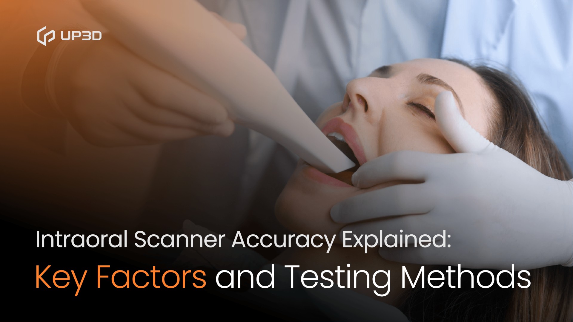
Accuracy is one of the most important performance indicators of any intraoral scanner. Whether you are capturing a single-crown preparation or a full-arch case, even a small deviation can affect restoration fit, occlusion, chairside time, and patient comfort. Yet “accuracy” is often used loosely, and many clinicians are unsure what it truly means or how it is measured.
This guide breaks down what scanner accuracy actually includes, why it varies between devices, and how accuracy is tested in dental research—so you can make better decisions when evaluating scanners or upgrading your digital workflow.
What “Accuracy” Really Means in Intraoral Scanning
In dentistry, accuracy is defined using two technical metrics:
1. Trueness
How close the scan is to the real geometry of the teeth or model.
Higher trueness = the scanner captures details more faithfully.
2. Precision
How consistent the scanner is when repeating the same scan multiple times.
Higher precision = less variation between scans.
A scanner must perform well in both to be considered high-accuracy.
Key Factors That Affect Scanner Accuracy
Several engineering and workflow elements determine how accurate a scanner can be in real clinical use.
1. Optical System Design
The quality of the cameras and lenses directly affects detail capture.
Systems with larger depth of field, high-resolution sensors, and optimized illumination can scan:
-
Deep margins
-
Reflective surfaces
-
Challenging anatomy
Modern scanners often combine multiple imaging technologies to maintain accuracy even when scanning quickly.
2. Software Algorithms
Optical data alone is not enough. Accuracy is heavily influenced by the scanner's processing engine—especially:
-
Image stitching algorithms
-
AI-based noise removal
-
Surface reconstruction models
-
Color and texture calibration
Algorithms determine how well the scanner aligns images without distortion, particularly in full-arch scans where cumulative stitching errors are common.
3. Scanning Strategy
Even the best scanner can lose accuracy with poor technique.
Common factors include:
-
Angle and distance of scanning
-
Overlapping scan paths
-
Whether the operator follows a validated scanning sequence
-
Moisture control and soft-tissue movement
A stable scanning strategy results in more predictable outcomes.
4. Patient-Related Conditions
Accuracy decreases when there is:
-
Excess saliva or fogging
-
Limited space in posterior regions
-
Mobility of soft tissues
-
Strong reflections from metal restorations
This is why many scanners include fog-free tips or adaptive exposure adjustments.
5. Calibration and Hardware Stability
Over time, small shifts in sensors or optics may affect accuracy.
Using a scanner with:
-
Regular calibration reminders
-
Stable mechanical design
-
High-quality internal optics
helps sustain accuracy long-term.
How Accuracy Is Tested in Research
Accuracy evaluation follows standardized testing approaches used in clinical studies and product validation.
1. Lab Model Comparison (Most Common Method)
A reference model is scanned with a high-precision industrial scanner (usually <5 µm accuracy).
The intraoral scanner's output is then compared to this reference to calculate:
-
Deviations
-
Surface errors
-
Full-arch distortion patterns
This method is widely considered the gold standard.
2. In-Vivo Full-Arch Testing
Researchers scan real patients multiple times to evaluate precision and identify accuracy loss due to:
-
Movement
-
Moisture
-
Operator technique
This method better represents real clinical conditions.
3. Superimposition and Color Map Analysis
Two scan files are overlaid to visualize deviations.
Color maps highlight areas with:
-
High accuracy (green)
-
Positive deviation (yellow/red)
-
Negative deviation (blue)
This is the clearest way to evaluate clinical scanning quality.
Are All Scanners Equally Accurate Across Indications?
Not necessarily.
Some scanners excel in single-unit cases but struggle with full-arch accuracy. Others have strong algorithms that maintain uniform trueness across long spans.
Consider evaluating accuracy based on your primary indications:
-
Single crown / inlay / onlay: Most scanners meet clinical requirements
-
Implants: Requires high precision at the scanbody level
-
Full arch: Demands advanced stitching algorithms and stable optics
Understanding these differences helps ensure predictable outcomes.
A Quick Note on Modern AI-Enhanced Scanners
Today's newer scanners increasingly use AI for:
-
Noise suppression
-
Automatic margin refinement
-
Soft-tissue filtering
-
Enhanced stitching for full-arch scans
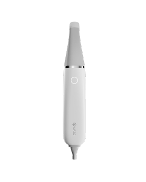
For example, scanners like the Clariscan UP610 from UP3D incorporate AI-driven processing to improve accuracy, especially in deep margins and long-span scans.
This type of subtle integration can support both clinical reliability and workflow efficiency—without compromising natural scanning speed.
Final Thoughts
Scanner accuracy is influenced by both engineering and clinical technique. Understanding trueness, precision, and the factors that affect them helps clinicians choose devices that deliver reliable outcomes across all indications.
As digital dentistry continues to evolve, accuracy will remain the foundation of predictable restorations—and a key requirement for every intraoral scanner in the modern workflow.