In collaboration with the Department of Prosthodontics, Victor Babeș University of Medicine and Pharmacy, Timișoara, Romania
Scanning a patient who is actively undergoing orthodontic treatment introduces a set of challenges that most intraoral scanners are not routinely tested against: metal brackets, archwires, ligatures, and the reflective surfaces they create. Add in a molar that needs endodontic retreatment and a chairside provisional crown, and you have a case that tests both the scanner and the clinical workflow.
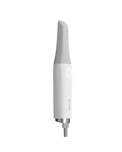
This case, completed at the Department of Prosthodontics at Victor Babeș University (UMFVBT) under the university's collaboration with UP3D, demonstrates how the Clariscan UP610 handled exactly that scenario — delivering accurate digital impressions through a full set of metal brackets, and feeding scan data into exocad for a chairside CAD/CAM provisional crown.
Patient Presentation

The patient, a 16-year-old male, presented with an asymptomatic periapical lesion on tooth 36 (lower left first molar), which had been previously endodontically treated. He was already undergoing orthodontic treatment on the maxillary arch, with mandibular brackets planned after the prosthodontic restoration was completed.
The treatment plan included:
-
Endodontic retreatment of tooth 36
-
Fiber post restoration
-
Digital impression with the UP610 for a chairside CAD/CAM long-term provisional crown
-
Definitive crown restoration to be completed after orthodontic treatment concludes
The decision to place a long-term provisional rather than a definitive crown was deliberate: the upcoming orthodontic treatment on the mandibular arch would alter the patient's occlusal relationships, making a final restoration premature. A reinforced composite provisional would protect the tooth during the orthodontic phase while preserving the option for an optimized definitive crown once the bite stabilized.

Pre-operative view of tooth 36 with existing restoration (left) and periapical radiograph showing the root canal filling and periapical radiolucency (right).
Digital Impression: Scanning Through Metal Brackets
After completing the endodontic retreatment and fiber post placement, the clinical team used the UP610 to capture digital impressions of both arches for the provisional crown design.
This is where the case became a real-world test of the UP610's orthodontic scanning capability. The maxillary arch was fully bracketed with metal brackets and archwire, while the mandibular arch presented the prepared tooth 36 alongside adjacent teeth. The scanner needed to handle two very different surface types in the same session: the highly reflective metal hardware on the upper arch, and the prepared tooth structure on the lower arch.
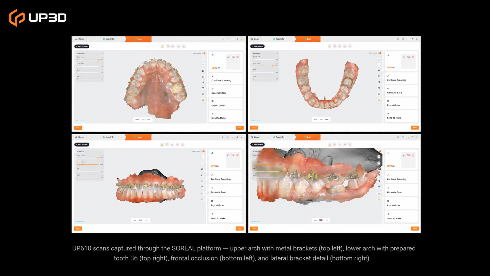
UP610 scans captured through the SOREAL platform — upper arch with metal brackets (top left), lower arch with prepared tooth 36 (top right), frontal occlusion (bottom left), and lateral detail showing bracket and archwire capture (bottom right).
The UP610 captured the metal brackets and archwire with full detail — no artifacts, no dropout areas around the reflective metal surfaces. The true-color rendering preserved the visual distinction between bracket metal, enamel, composite, and gingival tissue, giving the clinician an immediate on-screen verification that the scan was complete and accurate.
A closer look at the prepared tooth 36 confirmed that the fiber post area and remaining tooth structure were captured with sufficient detail for crown design — including the margin line and the transition from tooth structure to the post restoration.

Close-up scans of prepared tooth 36 — occlusal view showing the fiber post area (left) and lateral view showing margin detail (right).
Crown Design in exocad
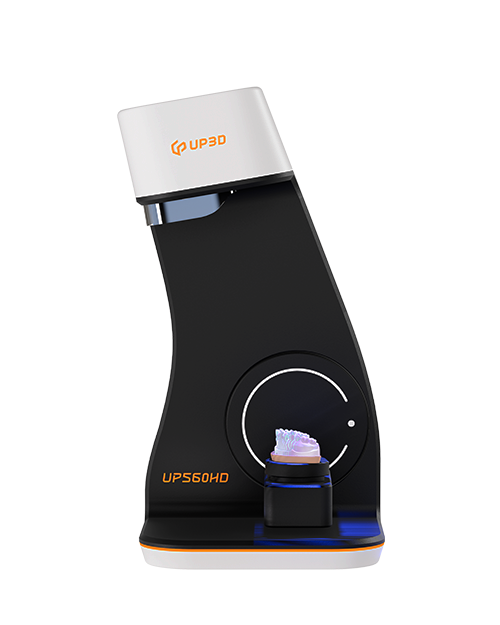
The scan data was exported from the SOREAL platform and imported into exocad for crown design. This step is worth noting: the UP610's open file format output (STL, PLY, OBJ) allows seamless integration with third-party CAD software — clinicians and labs are not locked into a single ecosystem.
In exocad, the provisional crown was designed with full anatomic contours, with the software's antagonist analysis verifying the occlusal relationship against the bracketed upper arch. The design was finalized and sent to the milling unit for chairside fabrication.
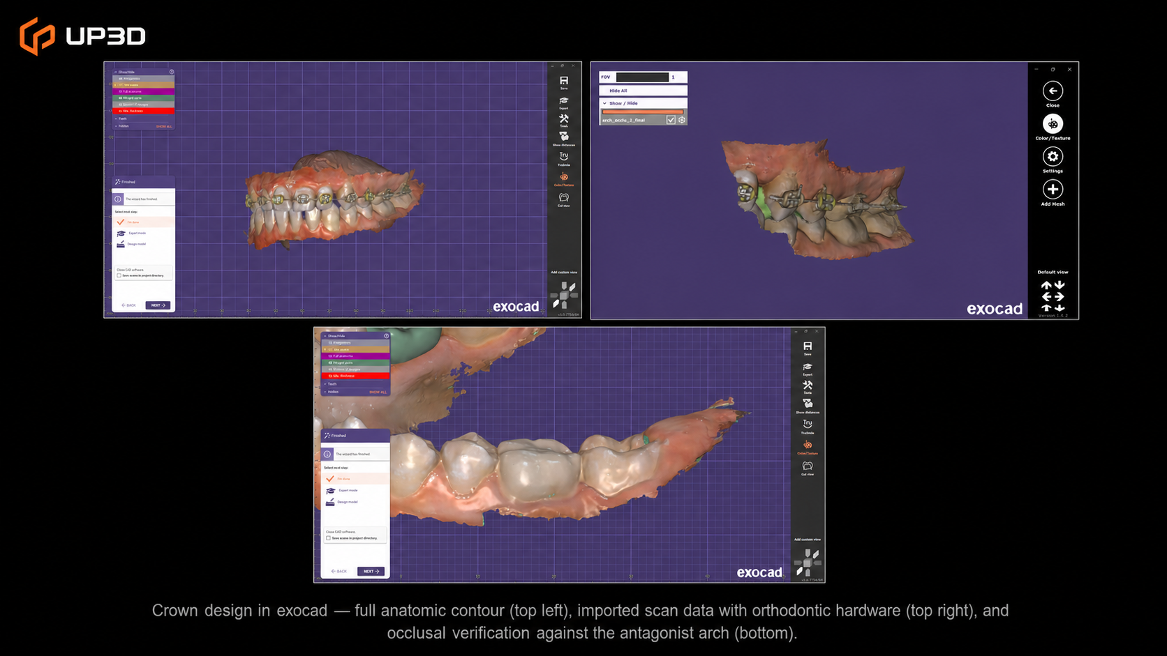
Crown design in exocad — full anatomic contour (left), imported scan data with orthodontic hardware visible (center), and occlusal verification against the antagonist arch (right).
Chairside Fabrication and Cementation
The provisional crown was milled chairside from a BRILLIANT Crios block (Coltene) — a reinforced composite material designed for long-term provisional and semi-permanent restorations. This material choice aligned with the clinical plan: strong enough to protect the tooth during the orthodontic phase, while remaining easy to remove and replace when the definitive restoration is placed.
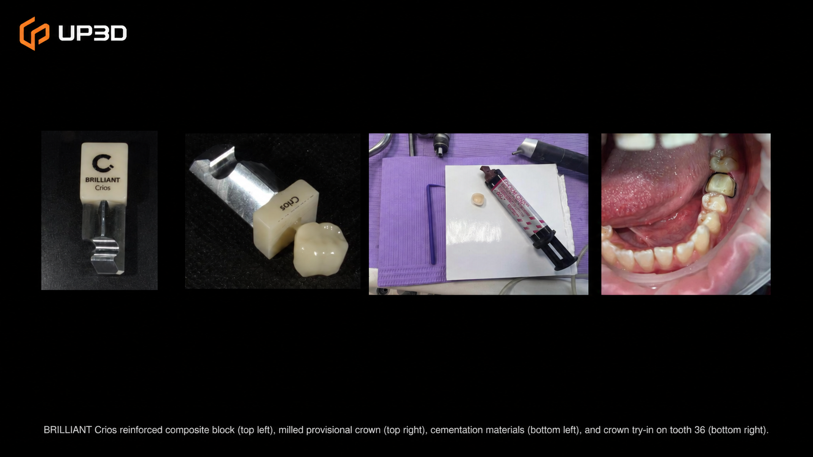
BRILLIANT Crios reinforced composite block (top left), milled provisional crown (top right), cementation materials (bottom left), and crown try-in on tooth 36 (bottom right).
The crown was cemented using G-CEM ONE (GC) self-adhesive protocol under rubber dam isolation — the same cementation approach used in Case 1, maintaining consistency across the department's digital workflow.
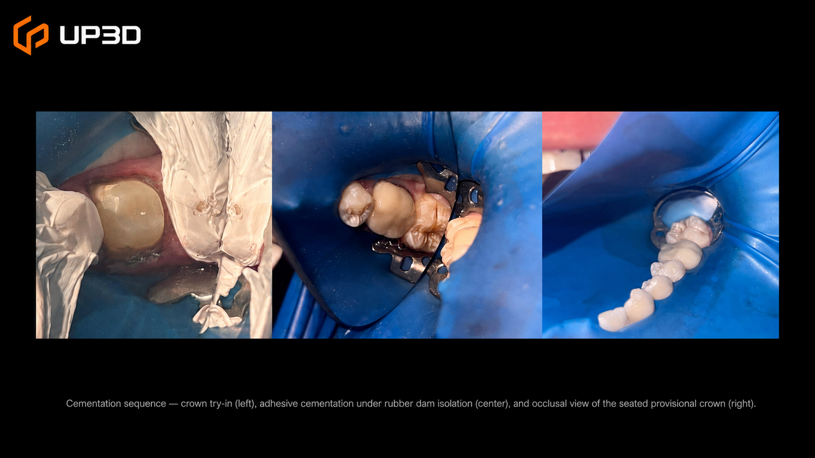
Cementation sequence — crown try-in (left), adhesive cementation under rubber dam isolation (center), and occlusal view of the seated provisional crown (right).
Clinical Outcome
The long-term provisional crown on tooth 36 achieved a natural integration with the surrounding dentition. The occlusal anatomy milled from the exocad design provided functional contacts, while the BRILLIANT Crios material delivered an acceptable aesthetic match for the provisional phase.
Following the prosthodontic restoration, mandibular orthodontic brackets were placed — confirming that the provisional crown's design and material allowed for bracket bonding on adjacent teeth without interference. The patient will continue orthodontic treatment, with the definitive crown planned once the occlusion has been finalized.
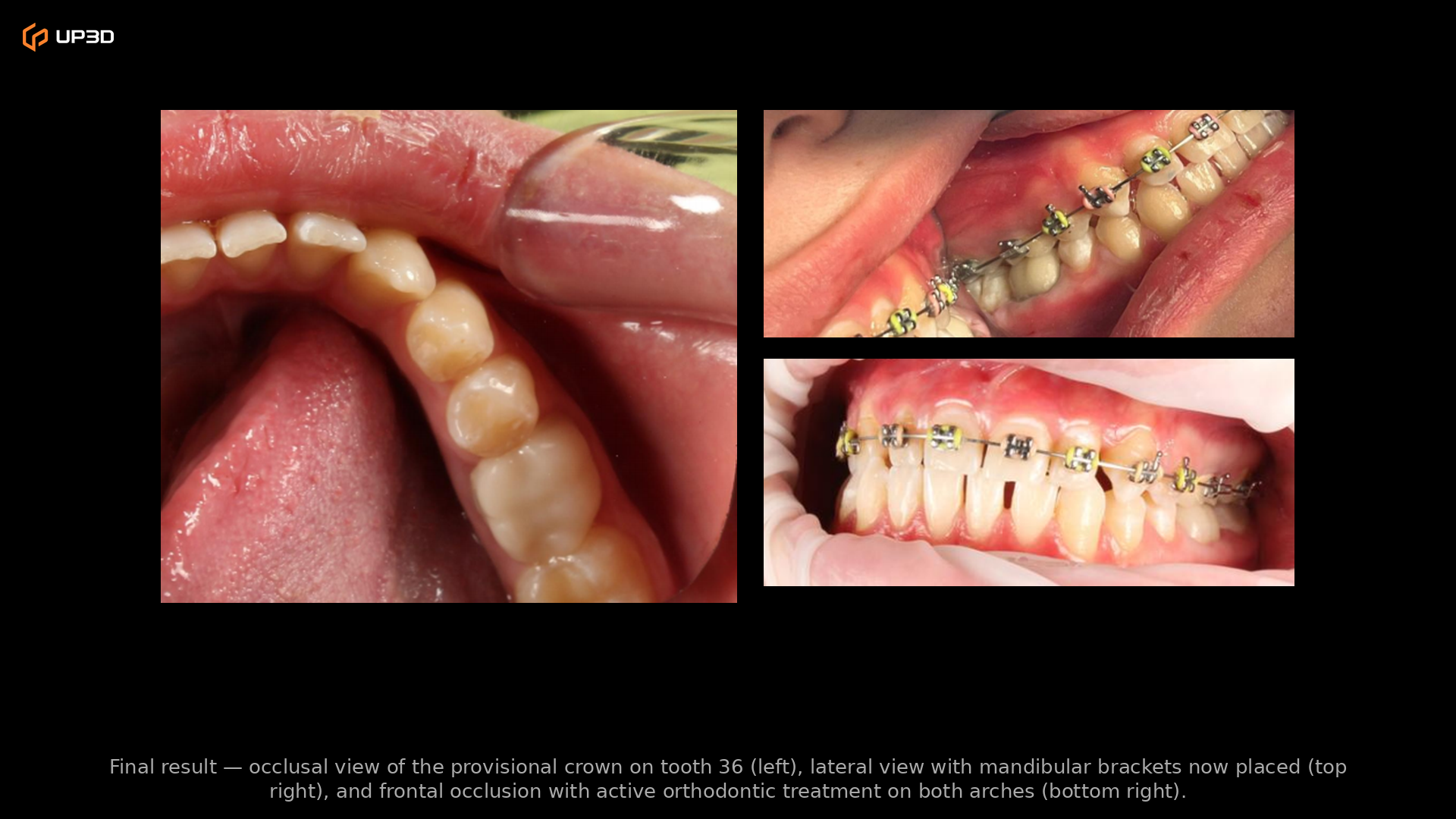
Final result — occlusal view of the provisional crown on tooth 36 (left), lateral view with mandibular brackets now placed (top right), and frontal occlusion showing the restoration integrated with active orthodontic treatment on both arches (bottom right).
Key Takeaways for Practitioners
Metal brackets are not a barrier. The UP610 captured metal brackets, archwires, and ligatures without artifacts or scan dropouts. For practices that see orthodontic patients needing restorative work, this eliminates the concern that digital impressions won't work around metal hardware.
Clinical decision-making drives material choice. A long-term provisional was the right call here — not because a definitive crown couldn't be fabricated, but because the patient's occlusion would change during orthodontic treatment. The digital workflow made it easy to scan, design, and mill a provisional chairside, with the option to repeat the process for the definitive crown later.
Open format means open choices. The scan data moved from the SOREAL platform to exocad without friction. Clinicians and labs using third-party CAD software can work with UP610 scan data (STL, PLY, OBJ) in their existing design environment — no ecosystem lock-in.
Two cases, one consistent workflow. This is the second clinical case from the UP3D-UMFVBT collaboration, following a post and core with zirconia crown case published earlier. Across both cases, the UP610 and SOREAL platform delivered a consistent, reliable scanning and data management workflow — across different patient ages, clinical scenarios, and restoration types.
About This Collaboration
This clinical case is part of an ongoing academic collaboration between UP3D and the Department of Prosthodontics at Victor Babeș University of Medicine and Pharmacy (UMFVBT) in Timișoara, Romania. The partnership integrates the Clariscan UP610 into the department's clinical and educational activities, generating real-world evidence of digital scanning performance across a range of prosthodontic applications.
Interested in seeing how the UP610 performs in your practice? Contact UP3D to learn more or request a demo.
Clinical images courtesy of the Department of Prosthodontics, UMFVBT. Patient consent obtained. All patient-identifiable information has been anonymized.